Scanning Electron Microscope
To operate SEM and the attached analytical instruments, you must pass both written and hands-on tests. Become a qualified user.
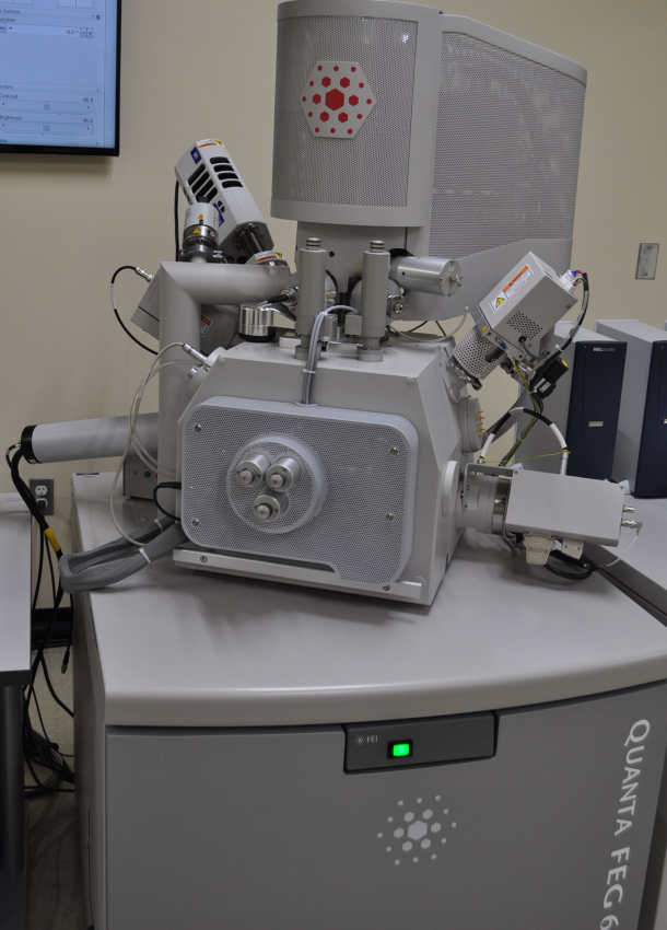
FEI Quanta FEG 650
The field-emission scanning electron microscope (FE-SEM) has multiple capabilities. Besides the traditional high vacuum mode to image conductive material, the FE-SEM has a low vacuum mode to image dry insulator and an environmental mode to image wet/biological samples without long hours of chemical fixation, which is prone to artifacts. The backscattered electron detector (BSED) can identify heavy/light elements. The STEM detector can image the internal structure of electron transparent materials with a maximum electron energy of 30 KeV. Stable and high current density of the FE-SEM can be used for e-beam lithography. Remote control software provides SEM access to classroom demonstration and research in regional campuses.
Oxford EDS with ULTIM MAX ∞ 100 Detector and EBSD with C SWIFT+ Detector
Analytical instruments attached to the SEM include an energy dispersive X-ray spectrometer (EDS), which provides information about chemical composition of a sample. Electron backscatter diffraction (EBSD) provides crystal structure information including crystal orientation and grain size distribution.
Publication Guidelines
In your publication, if the image, spectra or analysis is produced from the microscope at MCF, please add the following acknowledgment: “We acknowledge the support from the Microscopy Core Facility at Utah State University for the SEM result.” If any image in the publication was taken by the staff, please acknowledge the person properly.
Reservation Policy
- Available times: 11:00 AM to 5:00 PM from Monday to Thursday per week except for holidays. The minimum sign-up time is 1 hour. Usage exceeding the booked time will be charged in units of 15 minutes. Cancellation is acceptable by 5 PM, the day before the appointment. Otherwise, the user will be charged based on the reserved time.
- Instruments may be reserved up to two weeks in advance.
- Hydrocarbons accumulated in the chamber will reduce image quality. Samples should never have any kind of grease or loose particles on the surface. Please sonicate your samples with an acetone bath or isopropanol (if sample can’t tolerate acetone) prior to the SEM session and always wear clean gloves when handling samples.
- In the description box, please describe your sample name, chemical compositon, detector(s), vacuum mode, and SEM information you want to get.